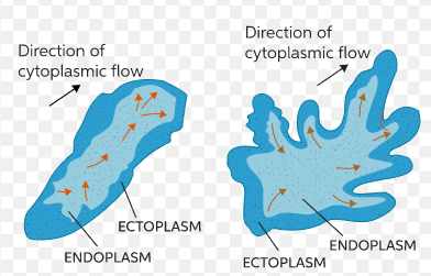
Amoeba is a type of single-celled organism that belongs to the kingdom Protista. It is known for its ability to move and change its shape, a characteristic known as an amoeboid movement. This movement results from the organism’s ability to extend and retract pseudopodia, temporary cell membrane extensions that function as legs.
“Sol-Gel” Theory of Locomotion in Amoeba
Amoeba is a primitive organism and lacks a specialized organ for locomotion, but it can still move towards favorable conditions, such as food, light, and water, or away from unfavorable conditions, such as toxins or high temperatures. The movement of amoeba is achieved through the formation of pseudopodia and the movement of cytoplasm within the cell.
Pseudopodia Formation
The formation of pseudopodia is a complex process involving the rearrangement of the actin filaments and microtubules within the cytoplasm. Actin filaments form a mesh-like structure at the cell’s leading edge, which acts as a scaffold for the pseudopodium formation. Microtubules play a role in shaping the pseudopodium and maintaining its structure.
The cytoplasm in the region of the pseudopodia is subjected to a concentration of actin-associated proteins, which causes the cytoplasm to become more fluid. This fluidity allows the cytoplasm to extend into the pseudopodium and pushes the cell membrane ahead.
Cytoplasmic Streaming
Cytoplasmic streaming is the movement of the cytoplasm within the cell, which helps to maintain the formation of the pseudopodia. It is driven by the beating of the microfilaments, similar to actin filaments but smaller in size.
The microfilaments beat in a coordinated manner, resulting in the movement of the cytoplasm in a specific direction. This movement helps to maintain the pseudopodium and provides the necessary energy for the organism to move.
Locomotion
The movement of amoeba is achieved by the extension and retraction of pseudopodia. When the organism wants to move in a particular direction, it extends a pseudopodium in that direction, which makes contact with the surface. The cytoplasm then moves into the pseudopodium, causing it to extend further and allowing the cell to move.
The retraction of the pseudopodium is achieved through the deformation of the actin filaments and the reformation of the microtubules. This process causes the pseudopodium to shrink, allowing the organism to change its shape and move in a different direction.
Factors affecting Locomotion
The movement of amoeba is influenced by several factors, including temperature, the presence of food or other stimuli, and the mechanical properties of the surface.
Temperature: Amoeba is a thermophilic organism which is sensitive to temperature changes. It moves faster in warmer temperatures and slower in cooler temperatures.
Food: The presence of food or other stimuli can also affect the movement of amoeba. The organism is able to sense the presence of food and move toward it. This type of movement is known as chemotaxis.
Surface Properties: The mechanical properties of the surface, such as roughness or stickiness, can also affect the movement of amoeba. The organism moves slower on rough surfaces or surfaces that are difficult to move on.
In conclusion, the amoeba is a single-celled organism capable of movement through the formation and movement of pseudopodia. This type of movement is driven by the rearrangement of actin filaments and microtubules and the beating of microfilaments within the cytoplasm.
The movement of amoeba is influenced by several factors, including temperature, the presence of food or other stimuli, and the mechanical properties of the surface. Understanding the mechanisms of amoeboid movement can provide valuable insight into cellular structure and function evolution.
In addition, the study of amoeba locomotion can have practical applications in fields such as medicine, as some pathogenic amoebae use their amoeboid movement to invade host tissues and cause disease. By understanding the mechanisms of amoeboid movement, researchers may be able to develop new strategies for preventing and treating amoebic infections.
In conclusion, the study of locomotion in amoeba is an important area of research with both theoretical and practical implications. Further research in this field has the potential to uncover new insights into the evolution of cellular function and provide new strategies for addressing amoebic diseases.

Molecular Mechanism of Locomotion in Amoeba
The molecular mechanism of locomotion in amoeba is driven by the coordinated movement of proteins within the cell. The two main proteins involved in this process are actin and myosin.
Actin is a protein that forms a network of filaments within the cell’s cytoplasm. These filaments are constantly rearranged to generate the movement of the cell, including the formation of pseudopodia. Actin filaments at the cell’s leading edge form a mesh-like structure, which acts as a scaffold for the pseudopodium formation.
Myosin is a protein that functions as a motor protein, using energy from ATP to generate movement. In the case of amoeba, myosin helps to drive the movement of the cytoplasm, which in turn helps to maintain the formation of the pseudopodia.
In addition to actin and myosin, other proteins, such as ARP2/3 and formins, play essential roles in the molecular mechanism of amoeboid movement. ARP2/3 and formins regulate the formation and elongation of actin filaments, which are crucial for forming pseudopodia.
The molecular mechanism of locomotion in amoeba is also influenced by the presence of signaling pathways, such as the Ras pathway, which help coordinate the cell’s movement in response to external stimuli.
In conclusion, the molecular mechanism of locomotion in amoeba is driven by the coordinated movement of actin, myosin, and other proteins, as well as the presence of signaling pathways. The precise mechanisms of how these proteins and pathways interact to generate movement in amoeba is an area of active research. Further study in this field can uncover new insights into cellular function and locomotion.
What is amoeboid movement?
Amoeboid movement is a type of movement characterized by the formation and movement of pseudopodia, or temporary extensions of the cell membrane, which allows an organism to move in a directional manner. This movement is commonly seen in single-celled organisms such as amoebae and some white blood cells. The movement is driven by the rearrangement of actin filaments and microtubules within the cytoplasm. It is used by cells to explore their environment, find food, and move towards or away from stimuli. Amoeboid movement is a highly dynamic and flexible form of movement and is an essential feature of many single-celled organisms, including some that are medically important.
What are pseudopodia?
Pseudopodia (also known as pseudopods) are temporary, finger-like projections of the cell membrane that extend from the cell body and are used for movement and/or feeding. They are common in single-celled organisms, such as amoebae, and play a crucial role in the amoeboid movement. The movement of pseudopodia is driven by the coordinated rearrangement of actin filaments and microtubules within the cytoplasm. It helps cells to explore their environment, find food, and respond to stimuli.
Pseudopodia can have different shapes, including lobopodia (rounded pseudopodia), filopodia (thread-like pseudopodia), and axopodia (long, thin pseudopodia with microtubules at the core). The type of pseudopodium that is formed depends on the cell’s needs and the specific context in which it is found.
In addition to being necessary for movement and feeding, pseudopodia also play essential roles in other cellular processes, such as phagocytosis (the uptake of particles by cells) and chemotaxis (the movement of cells in response to chemical gradients).
Types of pseudopodia in Amoeba
There are several different types of pseudopodia that can be formed by amoeba, including:
- Lobopodia: These are rounded pseudopodia commonly formed by amoeba and used for movement and feeding.
- Filopodia: These are thin, thread-like pseudopodia that are formed by the extension of actin filaments. They are typically used for exploratory movement and are essential for cell sensing and communication.
- Axopodia: These are long, thin pseudopodia with microtubules at their core. They are often found in amoeboid organisms that live in aquatic environments and play an essential role in helping the cells to maintain their shape and orientation.
- Reticulopodia: These are network-like pseudopodia that form a complex network of extensions from the cell body. They are commonly found in cells that are involved in phagocytosis or the uptake of particles.
The specific type of pseudopodium that is formed by an amoeba depends on its needs and the context in which it is found. For example, an amoeba searching for food may form filopodia to extend and explore its surroundings. If it is under attack by a predator, it may form reticulopodia to help it engulf and neutralize the threat.
Important Examination Questions on Locomotion in Amoeba
- Describe the molecular mechanism that drives locomotion in amoeba.
- How does the formation and movement of pseudopodia contribute to amoeboid movement?
- Explain the role of actin and myosin in the movement of amoeba.
- Discuss the influence of environmental factors, such as temperature and the presence of food, on the movement of amoeba.
- Describe the different types of pseudopodia that can be formed by amoeba, and give examples of their different functions.
- How does the process of amoeboid movement compare to other forms of cellular movement, such as ciliary and flagellar movement?
- Discuss the practical implications of the study of locomotion in amoeba, including its potential applications in fields such as medicine.
- Explain the role of signaling pathways, such as the Ras pathway, in regulating amoeboid movement.
- How has the study of amoeboid movement contributed to our understanding of cellular structure and function?
- What future directions do you see for research on the topic of amoeboid movement and its role in cellular biology?