Discover the intricate and vital role of the canal system in sponges. This blog post provides a comprehensive overview of the definition, histology, types, and functions of the canal system in sponges. Learn how it supports water filtration, circulation, respiration, defense against predators, and reproduction, and discover how it plays a crucial role in their survival and metabolism.
The canal system in sponges is a complex and essential feature that plays a critical role in the survival and metabolism of these aquatic animals. Sponges belong to the phylum Porifera and are known for their unique and specialized structures, including the canal system. This blog post provides a comprehensive overview of the canal system in sponges, including its definition, histology, types, and functions.
Definition of Canal System
The canal system in sponges is a series of interconnected canals and chambers that run through the sponge’s body. These canals are lined with specialized cells called choanocytes, which significantly filter water and capture food particles.
Histology of Canal System
Sponges are composed of two main cell types, choanocytes, and porocytes. Choanocytes line the walls of the canal system and have flagella that beat to create a flow of water through the sponge. Porocytes are specialized cells forming pores in the sponge’s body wall, allowing water to enter and exit the canal system.
Histology of Canal System
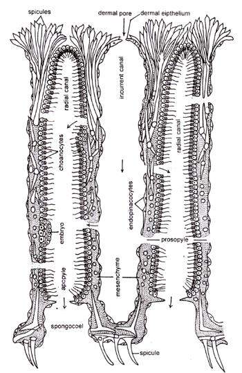
The various microscopic components that makeup Scypha’s body wall are referred to as histology components. Cell elements, skeletal elements, and mesenchymal substances are the three categories into which the elements can be classified.
With the exception of choanocytes, the cells of sponges appear to be just modified versions of undifferentiated amoeboid cells, which are analogous to the primordial connective tissue cell of higher animals.
The nucleus is located in a thickened central bulge of each pinacocyte. The incurrent canals and spongocoel are lined with pinacocytes (endopinacocytes). The cells lining the current canals are known as skeletogenous cells and are very contractile.
Ectodermal cells
These cells have been flattened into scales, have discrete nuclei, and have edges that are tightly cemented together to form an epithelium. Pinacocytes are the cells that make up the dermal layer that covers the sponge’s outside.
Endodermal cells
Endodermal cells, referred to as choanocytes, are only found in the radial canals.
Myocytes
These fusiform contractile cells are often organized in a circle to form sphincters. Prosopyles and apopyles help to close the apertures.
Mesenchyme Substance
Mesenchyme Substance The animal gains rigidity as a result. There are some amoebocytes or free-moving cells in the mesogloea.

Types of Canal System
Sponges can be classified into two main types based on the structure of their canal system: Asconoid, Syconoid, and Leuconoid.
Ascon Type of Canal System
The simplest of the three canal systems is this one. It can be found in asconoid sponges like Leucosolenia and some of the second sponges’ developmental phases.
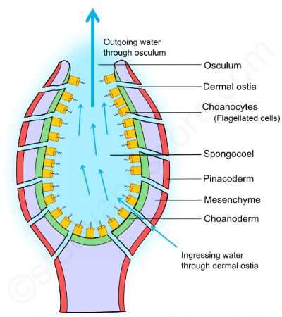
The asconoid sponges have numerous tiny openings on their body surface known as incurrent pores or Ostia. The tube-like cells known as porocytes contain these pores as intracellular gaps. These pores enter directly into the spongocoel and extend radially into the mesenchyme.
The sponge’s spongocoel is its single largest and most roomy cavity. Choanocytes, or flattened collar cells, line the spongocoel. The distal end of the sponge cool has an osculum, a tiny circular hole that opens to the exterior and is fringed with numerous monaxon spicules.
Through the Ostia, the surrounding seawater enters the network of canals. The beating of the collar cells’ flagella helps to keep the water flowing. The enormous spongocoel contains a lot of water that can’t be squeezed out by a single osculum; hence the water flow is slow.
The course of water current in Asconoid type canal system
Ingressing water>> Ostia >> Spongocoel>> Osculum >> outside

Sycon Type of Canal System
Comparatively speaking, the ascon canal system is simpler than the Sycon type. Syconoid sponges like Scypha have this particular form of channel system as a distinguishing feature. Theoretically, this canal system can be created by folding the walls of an asconoid in a horizontal direction. Additionally, Scypha’s embryonic development vividly demonstrates how the asconoid pattern changes into the Syconoid pattern.
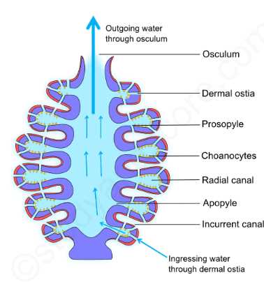
The radial and incurrent canals, which run parallel to and alternate with one another, are two types of canals found in the body walls of Syconoid sponges. Despite having a blind end into the body wall, these canals are connected via tiny pores. On the body’s surface are incurrent pores, also referred to as dermal Ostia. Incurrent canals are entered by these incurrent pores.
Since pinacocytes rather than choanocytes line the incurrent canals, they are non-flagellated. Through the tiny holes known as Prosopyles, incurrent canals enter neighboring radial canals. Radial canals, on the other hand, are flagellated because choanocytes line them. Internal Ostia or apopyles allow these canals to enter the central spongocoel.
A spongocoel is a small, non-flagellated chamber lined by pinacocytes in the Sycon type of canal system. It has an excurrent aperture called an osculum that connects to the outside world and is similar to the ascon type of canal system.
Few species, such as Grantia, whose incurrent canals are irregular and branching to produce huge sub-dermal areas, exhibit a more sophisticated type of the Sycon canal system. This results from the cortex’s growth, which involved pinacoderm and mesenchyme spreading throughout the sponge’s entire outer surface.
The course of water current in Asconoid type canal system
Ingressing water >> dermal ostia >> incurrent canal >> Prosopyles >> Radial canals >> Apopyles >> Spongocoel >> Osculum >> Outside

Leucon Type of Canal System
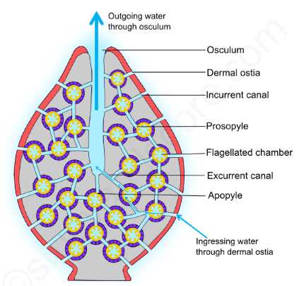
This form of canal system develops due to the body wall of the Sycon type of canal system becoming further folded. The Leuconoid variety of sponges, including Spongilla, have a unique canal system. Due to the complexity of the canal system, radial symmetry is lost in this form, leading to irregular symmetry.
Comparatively speaking to the asconoid and Syconoid types, the flagellated chambers are tiny. These sphere-shaped chambers are lined with choanocytes. Pinacocytes line all other spaces. Through Prosopyles, the incurrent canals enter flagellated chambers. Through apopyles, these flagellated chambers converse with the excurrent canals. Excurrent canals form as a result of spongocoel division and shrinking. This canal system lacks the wide, open spongocoel found in Asconoid and Syconoid canal systems. The spongocoel is significantly diminished here. Through the osculum, this excurrent canal finally connects with the outer world.

In its evolutionary pattern, the Leucon type of canal system has the following three successive grades:
Eurypylous type
The Leuconoid canal system is classified as having a eurypylous type, the simplest and most basic type. Through large openings known as apopyles, the flagellated chambers of this type immediately interface with the excurrent canal. Example: Plakina
Aphodal type: The apopyles are dragged out into a small canal called an aphodas in this type of canal system. This links the excurrent canals to the flagellated chambers. Example: Geodia
Diplodal type: Between the incurrent canal and the flagellated chamber in some sponges, there is also a second, tiny tube called a prosodus. This configuration results in a canal system of the diplodal type. Ex: Spongilla
Functions of Canal System
The canal system in sponges plays several important roles, including:
- Water filtration: The canal system filters water, removes food particles captured by choanocytes, and transfers them to other cells in the sponge for digestion.
- Circulation: The flow of water through the canal system helps distribute nutrients and oxygen throughout the sponge’s body.
- Excretion: The canal system also plays a role in removing waste products from the sponge and maintaining a healthy balance of ions and other substances in the water.
- Protection: The flow of water through the canal system also helps to protect the sponge from predators by creating a current that disrupts the movement of potential predators.
In conclusion, the canal system in sponges is a critical component of their anatomy and plays multiple essential roles in their survival and metabolism. Understanding the structure and function of the canal system is essential for understanding the biology of sponges and their role in marine ecosystems.
The canal system of sponges is also responsible for regulating water flow through the sponge. The flow of water is created by beating the choanocytes’ flagella, which creates a current that circulates water and helps maintain a constant flow through the canal system. Water flow also helps remove waste products from the sponge and maintains a healthy balance of ions and other substances in the water.
Sponges have a unique form of respiration that involves the exchange of gases through the choanocytes and other cells in the canal system. Oxygen is taken in from the water, while carbon dioxide and other waste products are expelled into the water. This type of respiration allows sponges to remain in constant contact with their watery environment and ensures a steady supply of oxygen and the removal of waste products.
In addition to filtering water and removing food particles, the choanocytes in the canal system also play a role in defense of the sponge. The flagella movement creates a current that helps to disrupt the activity of potential predators and deter them from attacking the sponge. In some species of sponges, the choanocytes can also release toxins or other chemical compounds that can deter predators.
Sponges also have the ability to reproduce both sexually and asexually. During sexual reproduction, gametes are produced and released into the water, where they can fuse to form a zygote. In asexual reproduction, the sponge can produce buds that grow into new sponges. Both of these processes can occur within the canal system, and the new individuals can then grow and develop into mature sponges.
In summary, the canal system in sponges is a complex and essential feature that plays a crucial role in their survival and metabolism. It helps filter water and capture food particles, circulate water and nutrients, regulate water flow, support respiration, defend against predators, and facilitate reproduction. Understanding the canal system and its functions is important for understanding the biology of sponges and their place in marine ecosystems.